Diagnosis of Osgood Schlatters Disease
How to diagnose Osgood Schlatters Disease?Clinical examination is usually all that is required as the enlarged lump and ‘Ouch’ reaction of the patient is pretty indicative of Osgood Schlatters Disease. Plus the age of the patient and history of the problem must match the classic group. However there are some more serious conditions such as infection or tumours that may need to be excluded if there is any uncertainty, and parents or carers of children with knee pain should always err on the side of caution and get a diagnosis from a qualified health practitioner such as a Medical Doctor or Physiotherapist/ Athletic Trainer.
Medical imaging tests may be needed to accurately diagnose the condition and its staging (severity).
X-rays have been traditionally used and show a clear separation of fragments of the tibial tubercle
More recently ultra-sound scans can also show the level of tendon inflammation and as these are less dangerous for using around growth plates on children, they are often preferred to X-rays.
Ultra-sound scan showing elevated bony fragment (courtesy of University of Greenwich, UK)
MRI scans show both of the above but are more expensive and time consuming to perform.
CT scans can also be very informative but these are not as easily available.

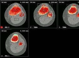
pQCT scan showing detached bony fragment at top of tibia (courtesy of Novotec Medical Ltd, Germany)
